

What Organelle Is In Plant Cells But Not Animals
Learning Outcomes
- Place key organelles present simply in plant cells, including chloroplasts and central vacuoles
- Identify key organelles present only in brute cells, including centrosomes and lysosomes
At this point, it should be clear that eukaryotic cells take a more circuitous structure than exercise prokaryotic cells. Organelles allow for various functions to occur in the cell at the same time. Despite their primal similarities, there are some striking differences between fauna and plant cells (see Figure 1).
Animal cells have centrosomes (or a pair of centrioles), and lysosomes, whereas plant cells practice not. Establish cells take a cell wall, chloroplasts, plasmodesmata, and plastids used for storage, and a large central vacuole, whereas animal cells do not.
Practice Question

Effigy 1. (a) A typical animal jail cell and (b) a typical plant prison cell.
What structures does a plant cell have that an fauna cell does not have? What structures does an animal cell have that a institute cell does not have?
Show Answer
Plant cells have plasmodesmata, a cell wall, a large central vacuole, chloroplasts, and plastids. Beast cells have lysosomes and centrosomes.
Found Cells
The Cell Wall
In Figure 1b, the diagram of a plant cell, you see a structure external to the plasma membrane called the cell wall. The prison cell wall is a rigid covering that protects the cell, provides structural support, and gives shape to the cell. Fungal cells and some protist cells also have cell walls.
While the chief component of prokaryotic cell walls is peptidoglycan, the major organic molecule in the plant cell wall is cellulose (Effigy 2), a polysaccharide made up of long, straight chains of glucose units. When nutritional information refers to dietary fiber, it is referring to the cellulose content of food.

Figure 2. Cellulose is a long chain of β-glucose molecules connected by a 1–four linkage. The dashed lines at each end of the effigy point a series of many more glucose units. The size of the folio makes it impossible to portray an entire cellulose molecule.
Chloroplasts

Figure 3. This simplified diagram of a chloroplast shows the outer membrane, inner membrane, thylakoids, grana, and stroma.
Similar mitochondria, chloroplasts also have their own DNA and ribosomes. Chloroplasts function in photosynthesis and tin be found in photoautotrophic eukaryotic cells such as plants and algae. In photosynthesis, carbon dioxide, water, and light energy are used to make glucose and oxygen. This is the major difference betwixt plants and animals: Plants (autotrophs) are able to make their own food, like glucose, whereas animals (heterotrophs) must rely on other organisms for their organic compounds or nutrient source.
Like mitochondria, chloroplasts have outer and inner membranes, merely within the space enclosed by a chloroplast'southward inner membrane is a ready of interconnected and stacked, fluid-filled membrane sacs called thylakoids (Effigy three). Each stack of thylakoids is called a granum (plural = grana). The fluid enclosed by the inner membrane and surrounding the grana is called the stroma.
The chloroplasts incorporate a green pigment called chlorophyll, which captures the free energy of sunlight for photosynthesis. Similar found cells, photosynthetic protists also have chloroplasts. Some bacteria also perform photosynthesis, but they practise not have chloroplasts. Their photosynthetic pigments are located in the thylakoid membrane within the prison cell itself.
Endosymbiosis
We have mentioned that both mitochondria and chloroplasts contain Dna and ribosomes. Have you lot wondered why? Strong prove points to endosymbiosis as the explanation.
Symbiosis is a relationship in which organisms from two split up species live in close clan and typically showroom specific adaptations to each other. Endosymbiosis (endo-= within) is a relationship in which 1 organism lives inside the other. Endosymbiotic relationships abound in nature. Microbes that produce vitamin Chiliad live within the human gut. This relationship is beneficial for us because we are unable to synthesize vitamin K. It is likewise benign for the microbes because they are protected from other organisms and are provided a stable habitat and arable food by living within the big intestine.
Scientists accept long noticed that bacteria, mitochondria, and chloroplasts are similar in size. We also know that mitochondria and chloroplasts accept DNA and ribosomes, just as bacteria do. Scientists believe that host cells and bacteria formed a mutually beneficial endosymbiotic relationship when the host cells ingested aerobic bacteria and cyanobacteria but did not destroy them. Through evolution, these ingested bacteria became more specialized in their functions, with the aerobic bacteria condign mitochondria and the photosynthetic bacteria becoming chloroplasts.
Endeavor Information technology
The Fundamental Vacuole
Previously, nosotros mentioned vacuoles as essential components of institute cells. If you look at Figure 1b, you volition come across that plant cells each take a large, central vacuole that occupies most of the cell. The central vacuole plays a key role in regulating the cell's concentration of water in changing environmental conditions. In plant cells, the liquid inside the central vacuole provides turgor pressure, which is the outward pressure caused by the fluid inside the cell. Accept you e'er noticed that if you forget to water a plant for a few days, it wilts? That is because as the water concentration in the soil becomes lower than the h2o concentration in the establish, water moves out of the central vacuoles and cytoplasm and into the soil. As the central vacuole shrinks, it leaves the prison cell wall unsupported. This loss of support to the cell walls of a plant results in the wilted appearance. When the central vacuole is filled with water, information technology provides a low energy means for the institute jail cell to aggrandize (as opposed to expending energy to actually increase in size). Additionally, this fluid can deter herbivory since the biting taste of the wastes it contains discourages consumption past insects and animals. The primal vacuole also functions to store proteins in developing seed cells.
Beast Cells
Lysosomes

Figure 4. A macrophage has phagocytized a potentially pathogenic bacterium into a vesicle, which then fuses with a lysosome inside the cell so that the pathogen can exist destroyed. Other organelles are present in the jail cell, merely for simplicity, are not shown.
In animal cells, the lysosomes are the jail cell's "garbage disposal." Digestive enzymes within the lysosomes help the breakup of proteins, polysaccharides, lipids, nucleic acids, and even worn-out organelles. In single-celled eukaryotes, lysosomes are of import for digestion of the nutrient they ingest and the recycling of organelles. These enzymes are active at a much lower pH (more than acidic) than those located in the cytoplasm. Many reactions that take place in the cytoplasm could not occur at a low pH, thus the reward of compartmentalizing the eukaryotic cell into organelles is apparent.
Lysosomes as well use their hydrolytic enzymes to destroy affliction-causing organisms that might enter the prison cell. A skillful example of this occurs in a group of white blood cells chosen macrophages, which are part of your torso's immune system. In a process known as phagocytosis, a section of the plasma membrane of the macrophage invaginates (folds in) and engulfs a pathogen. The invaginated section, with the pathogen inside, then pinches itself off from the plasma membrane and becomes a vesicle. The vesicle fuses with a lysosome. The lysosome's hydrolytic enzymes and so destroy the pathogen (Figure four).
Extracellular Matrix of Creature Cells

Figure 5. The extracellular matrix consists of a network of substances secreted by cells.
Most creature cells release materials into the extracellular space. The primary components of these materials are glycoproteins and the protein collagen. Collectively, these materials are called the extracellular matrix (Figure 5). Not simply does the extracellular matrix concord the cells together to course a tissue, but it also allows the cells within the tissue to communicate with each other.
Claret clotting provides an example of the function of the extracellular matrix in cell communication. When the cells lining a blood vessel are damaged, they display a poly peptide receptor called tissue factor. When tissue factor binds with another factor in the extracellular matrix, information technology causes platelets to adhere to the wall of the damaged blood vessel, stimulates side by side smooth muscle cells in the claret vessel to contract (thus constricting the claret vessel), and initiates a series of steps that stimulate the platelets to produce clotting factors.
Intercellular Junctions
Cells can likewise communicate with each other by directly contact, referred to as intercellular junctions. There are some differences in the ways that plant and animal cells practise this. Plasmodesmata (singular = plasmodesma) are junctions between plant cells, whereas animal prison cell contacts include tight and gap junctions, and desmosomes.
In general, long stretches of the plasma membranes of neighboring plant cells cannot touch one some other because they are separated past the cell walls surrounding each prison cell. Plasmodesmata are numerous channels that pass between the cell walls of next plant cells, connecting their cytoplasm and enabling signal molecules and nutrients to be transported from cell to prison cell (Figure 6a).
A tight junction is a watertight seal between ii adjacent brute cells (Figure 6b). Proteins hold the cells tightly against each other. This tight adhesion prevents materials from leaking between the cells. Tight junctions are typically constitute in the epithelial tissue that lines internal organs and cavities, and composes most of the pare. For case, the tight junctions of the epithelial cells lining the urinary float forestall urine from leaking into the extracellular space.
Also found only in animal cells are desmosomes, which deed like spot welds between adjacent epithelial cells (Figure 6c). They keep cells together in a canvas-like germination in organs and tissues that stretch, like the skin, heart, and muscles.
Gap junctions in animal cells are similar plasmodesmata in plant cells in that they are channels betwixt adjacent cells that allow for the transport of ions, nutrients, and other substances that enable cells to communicate (Effigy 6d). Structurally, still, gap junctions and plasmodesmata differ.
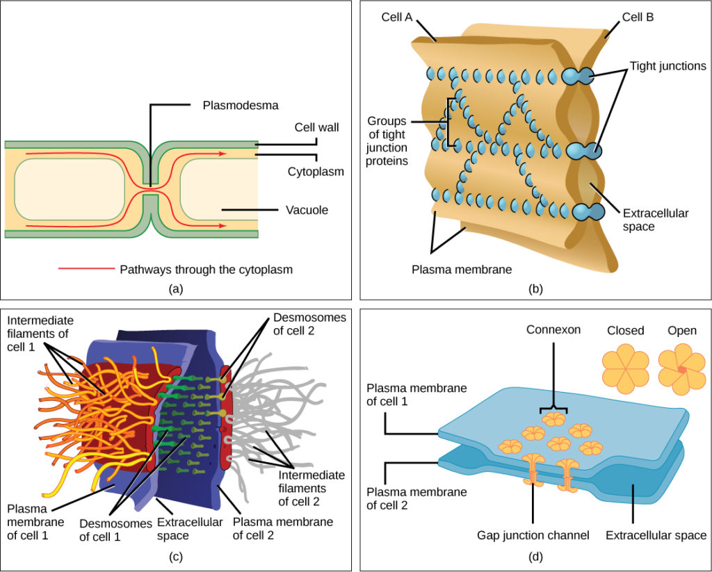
Figure six. There are four kinds of connections between cells. (a) A plasmodesma is a channel between the jail cell walls of two adjacent institute cells. (b) Tight junctions join adjacent fauna cells. (c) Desmosomes join ii animal cells together. (d) Gap junctions deed as channels between animal cells. (credit b, c, d: modification of work past Mariana Ruiz Villareal)
Contribute!
Did y'all have an idea for improving this content? We'd love your input.
Ameliorate this pageLearn More
Source: https://courses.lumenlearning.com/wm-nmbiology1/chapter/animal-cells-versus-plant-cells/
Posted by: ahrenssaisent.blogspot.com
0 Response to "What Organelle Is In Plant Cells But Not Animals"
Post a Comment